Back
 (Download Image)
The atomic-scale structure of the enzyme tied to the single-celled parasite responsible for African sleeping sickness.
(Download Image)
The atomic-scale structure of the enzyme tied to the single-celled parasite responsible for African sleeping sickness.
First-ever determination of protein structure with X-ray laser
(Download Image)
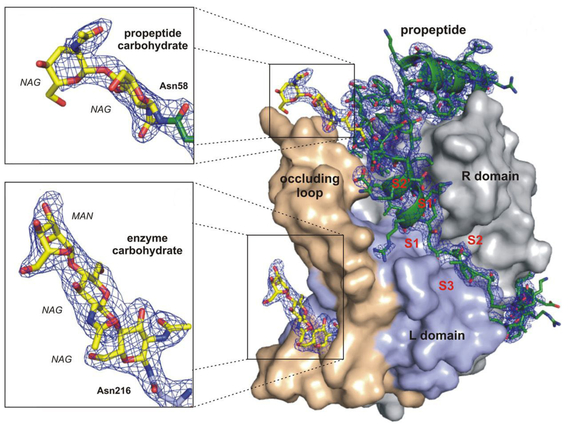
The atomic-scale structure of the enzyme tied to the single-celled parasite responsible for African sleeping sickness.
An international team of researchers, including LLNL physicist Matthias Frank and postdoc Mark Hunter, have for the first time used an ultra-intense X-ray laser to determine the previously unknown atomic-scale structure of a protein.
The work was reported in the online edition of Science , which also featured the story as a News Flash. The team determined the structure of an enzyme key to the survival of the single-celled parasite Trypanosoma brucei, responsible for African sleeping sickness, a disease that kills 30,000 people each year.
This new structural information should help guide the search for drugs that act like the propeptide, tying up the enzyme and killing the parasite. To determine the structure of the precursor form of the protein -- which does not form crystals large enough for traditional X-ray diffraction -- submicron nanocrystals produced by the parasite were analyzed by the "diffraction before destruction" technique, in which individual nanocrystals are passed, one by one, through the X-ray beam at the Linac Coherent Light Source, followed by "stacking"of the resultant diffraction data -- in this case, from 178,875 individual nanocrystals.
The achievement also demonstrates that the approach can provide otherwise unobtainable biomolecular information, potentially ushering in a new era of protein crystallography.
The research also was r ecognized as one of the "Top 10 2012 Science Breakthroughs of the Year" in Science Magazine . See the story .
Livermore researchers -- whose participation in the research is supported by the LDRD Program -- include the development of the nanoparticle injectors, setting up laser pump probe experiments, sample preparation, damage modeling and data acquisition at the LCLS. The figure shows the quality of the electron density (blue) in the calculated structure.
March 11, 2013
[email protected]
925-422-9799


Contact
Anne M Stark[email protected]
925-422-9799
Tags
Physical and Life SciencesFeatured Articles