Understanding effects of tissue-specific extracellular matrix molecules on brain cells
 (Download Image)
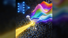
Illustration of electrodes on the multi-electrode array (MEA) detecting the communication (grey lines) between neurons within different neural network communities (colored circles). Greater cell-cell communication is observed in ECM-coated MEAs (MaxGel and bECM) compared to non-ECM coated MEA (PDL).
In vitro organ-on-a-chip experimental platforms hold great promise to predict human biology and human-relevant responses to chemical and biological stimuli. LLNL scientists and engineers are working to more faithfully recapitulate the brain microenvironment on multi-electrode arrays to study brain activity. In an article recently published in Scientific Reports, Livermore researchers describe the results of a study in which they examined the effects of tissue-like molecules, called extracellular matrix (ECM) molecules, on brain cell function.
Approximately 20% of the adult brain is composed of ECM molecules, molecules that contribute to the tissue architecture of the brain. Structurally, these molecules form a network that act as a physical barrier to reduce cell movement and the diffusion of proteins in the brain. The molecules also function to regulate a number of fundamental processes during brain development and can play a role in the healthy and diseased brain.
To more accurately mirror the tissue architecture of the brain, LLNL scientists extracted ECM from rat brain tissue, in addition to using a commercial human ECM source extracted human skin cultures (e.g., fibroblasts and epithelial cells) called MaxGel. The goal of this research was two-fold: (1) to determine whether increasing the complexity of ECM in the biological system improved brain activity, and (2) to understand whether a human-relevant ECM (and non-brain relevant ECM) can be used to increase the biological relevance of the system to mimic the brain in vivo.
This study highlights the general benefit of incorporating tissue-relevant ECM to augment the complexity of the biological system on the brain-on-a-chip platform, even if the ECM is not compositionally identical to brain ECM, as it can accelerate the ability of brain cells to communicate to one another and form neural networks without compromising the “speech patterns” or the intrinsic behavior of network connectivity. It is important to discover conditions (e.g., cellular complexity, tissue-specific ECM coating) that improve neuronal network formation and function, and in this research LLNL researchers were able to show that adding the tissue architecture of the brain is one modality and one step closer to mimicking properties of the brain in vivo.
This work was funded by the Laboratory Directed Research and Development Program (17-SI-002).
[D. Lam, H.A. Enright, J. Cadena, S.K.G. Peters, A.P. Sales, J. Osburn, D.A. Soscia, K.S. Kulp, E.K. Wheeler, and N.O. Fischer, Tissue-specific extracellular matrix accelerates the formation of neural networks and communities in a neuron-glia co-culture on a multi-electrode array, Scientific Reports 9, 4159 (2019), doi: 10.1038/s41598-019-40128-1]
Tags
Bioscience and BioengineeringBiosciences and Biotechnology
Physical and Life Sciences
Featured Articles