Back
 Previous
Next
Previous
Next
Seeing is believing: LLNL Postdoc reveals wonders of the protist world
(Download Image)
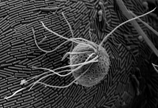
The oxymonad protist S accinobaculus (foreground, at about 12 microns in diameter) orbiting the much larger parabasalid protist Barbulanympha (background, at about 100 microns in diameter), which is covered in a dense monolayer of rod-shaped symbiotic bacteria. From the hindgut of the wood-feeding roach Cryptocercus . Images by Kevin J. Carpenter
(Download Image)
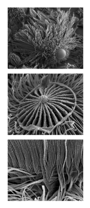
Trichonympha triptych , showing the termite hindgut protist Trichonympha (top: whole cell) and high resolution ultrastructural features (middle: Anterior "compressor fan" assembly organizing anterior flagella; bottom: Posterior view showing flagellar bundles).
No matter how sophisticated instruments have become, or how data intensive results may be, there is still something satisfying and frequently informative about seeing what a researcher is studying. Sometimes samples are so small, or so large, or so far away that it's almost impossible to get a visual perspective that makes any sense or reveals any insight.
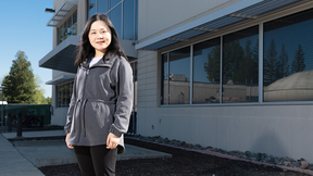
That's where the Lab's Kevin Carpenter comes in with his experience and expertise in sample preparation, microscopy, microanalysis and high resolution imaging.
"My parents took the family on frequent vacations to Yosemite and the California coast where I developed my lifelong interest in nature photography and biology," Carpenter said.
During his undergraduate study at UC Irvine, Carpenter developed an interest in the diversity of life and how the investigation of cellular structure and form can lead to and test hypotheses about evolution and ecology.
Combining his interests in organismal structure and photography led him to work in light and electron microscopy. He went on to develop and refine sample preparation, electron microscopy and imaging techniques on numerous projects including study of the evolution of pollen and leaf epidermal cell patterning of the earliest flowering plant lineages (e.g., water lilies) -- the topic of his dissertation research at UC Davis.
"After completing my dissertation, my interest in the diversity of life inspired me to turn my attention to a highly diverse group of microbes called protists which include protozoa and most species of algae," Carpenter said. "This led me to devise new sample preparation and electron microscopy techniques for imaging protists -- especially the highly complex and bizarre examples found in the guts of termites-during my first postdoc at the University of British Columbia. Very little scanning electron microscopy had ever been done on these organisms and our work revealed new cellular structures, new types of symbioses with bacteria, and also led to some interesting evolutionary and ecological hypotheses."
Becoming interested in the functional and ecological aspects of termite gut protists and their various symbioses with bacteria (some of which Carpenter's imaging revealed for the first time), he came to LLNL as a postdoc in 2009 to study these symbiotic relationships with the Lab's Cameca 50 NanoSIMS (high resolution imaging mass spectrometer) in the Chemical and Isotopic Signatures group in the Chemical Sciences Division. In 2010, Carpenter was awarded an LDRD (Labwide) grant to carry out his research.
"These protists are not only spectacular morphologically, but they also have biochemical and biophysical capabilities that make them very attractive for bioenergy research -- specifically the ability to efficiently break down wood into simpler carbon compounds and hydrogen gas. The application of NanoSIMS seemed like the next logical step for the study of these organisms. This instrument gives us very high resolution images showing distributions of elements and isotopes. It reveals how individual microbes are using and transferring isotopically labeled compounds we are introducing to the termites' diet and air," he said. Carpenter has further modified and adapted his methods for this research, and gave an invited seminar on his results at a meeting in Berlin, Germany in July 2011.
Since 2007, Carpenter's methods for imaging protists and their bacterial symbionts have led to nine peer reviewed articles and six journal covers, including the cover of December 2011 issue of Microbe . His micrographs also have been featured in textbooks, newspaper articles and on television programs. In 2010, the Microscopy Society of America awarded him a prestigious micrograph award for his collection of work. In addition to his scientific recognition, he also was an invited exhibitor of his work at shows held at two scientific meetings.
Jan. 17, 2012
[email protected]
Laboratory Directed Research and Development
Physical and Life Sciences
Contact
Brian D Johnson[email protected]
Tags
NanoSIMSLaboratory Directed Research and Development
Physical and Life Sciences
Featured Articles