Protein that prevents further cartilage damage
 (Download Image)
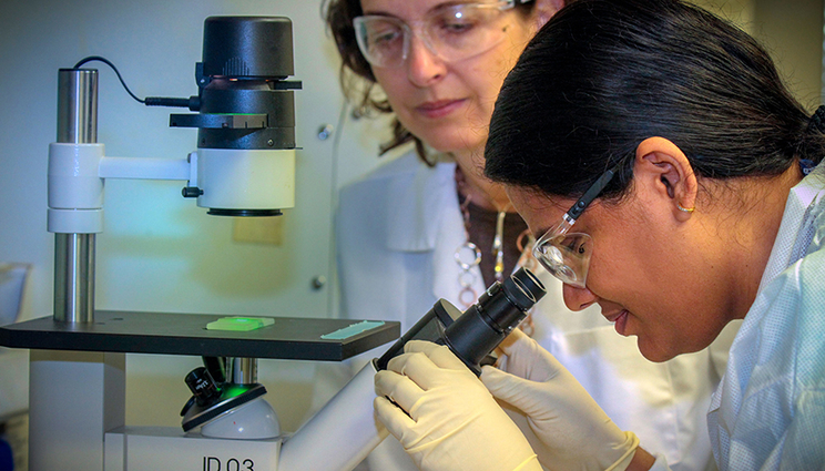
LLNL scientists Gaby Loots (left) and Aimy Sebastian count live cells for their research tying a specific protein to help prevent further cartilage damage in ACL patients.
(Download Image)
LLNL scientists Gaby Loots (left) and Aimy Sebastian count live cells for their research tying a specific protein to help prevent further cartilage damage in ACL patients.
There may be a protein in the body that hinders cartilage degradation in patients with a torn anterior cruciate ligament (ACL).
Lawrence Livermore National Laboratory (LLNL) scientists performed in vivo experiments on animal models of post traumatic osteoarthritis and found that sclerostin (a protein that in humans is encoded by the Sost gene), acts as a protective molecule immediately post joint injury to inhibit cartilage loss and joint calcification.
Patients with ACL ruptures are two times more likely to develop post traumatic osteoarthritis (PTOA). Annually, there are 900,000 knee injuries in the U.S, which account for 12 percent of all osteoarthritis (OA) cases. PTOA leads to reduced physical activity, deconditioning of the musculoskeletal system, and in severe cases, requires joint replacement to restore function.
"We discovered that administering sclerostin to injured joints would significantly slow down cartilage degradation post injury. The development of treatments that can be delivered to joints immediately post injury could provide attractive alternatives to surgery," said Gaby Loots, LLNL biologist and lead author of a study appearing in the Journal of Bone and Mineral Research.
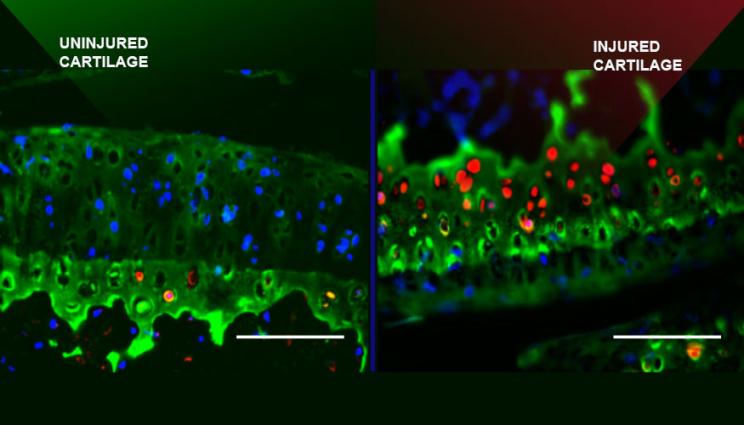
To determine whether elevated levels of sclerostin play a protective role in PTOA, the team examined the progression of OA using a noninvasive tibial compression overload model developed by Dr. Blaine Christiansen at UC Davis."We found that the transgenic mice overexpressing SOST develop moderate OA and display significantly less advanced PTOA phenotypes at 16 weeks post injury compared to the control mice," Loots said. In addition, the transgenic mice built approximately 65 percent less osteophytes (a bony outgrowth that aims to stabilize the injured joint) than the control group.
The increased risk of developing knee OA after injury to the ACL has been well documented both clinically and in experimental models. Clinical manifestation of PTOA is characterized by narrowing of the joint space, emergence of osteophytes, cartilage erosion and fibrillation
Biomechanical disturbances in the joint, such as lateral dislocation of the tibia, further the development of osteophytes in the lateral tibial-femoral compartment and cause misalignment, rotation and anterior dislocation of the joint; all these physical manifestations contribute to the emergence of intra-articular lesions. Cartilage lesions become further exacerbated through molecular changes in the joint, including the increase in the production of matrix-degrading enzymes, such as aggrecanases and metalloproteinases (MMPs).
Elevated levels of these enzymes enhance the loss of articular cartilage, increase the amount of pain experienced and lead to impaired joint mobility in more than 50 percent of individuals that sustained an ACL tear. In addition to changes in joint architecture and uneven biomechanical load distribution in the knee after an ACL tear, the individual can experience inflammatory responses, enzymatic cartilage destruction and osteophyte formation that will determine subsequent osteoarthritic results.
"Our results suggest that elevated levels of sclerostin, immediately post injury, can aid the joint in maintaining its articular cartilage integrity in PTOA," Loots said.
Other Livermore scientists include Jiun Chang, Deepa Murugesh, Aimy Sebastian, Nicholas Hum, Nicole Collette and Craig Blachette, as well as researchers from University of California Merced, University of California Davis Medical Center and Regenereron Pharmaceuticals.
Contact
 Anne M. Stark
[email protected]
(925) 422-9799
Related Links
Journal of Bone and Mineral ResearchTags
Physical and Life SciencesScience
Featured Articles