Back
 (Download Image)
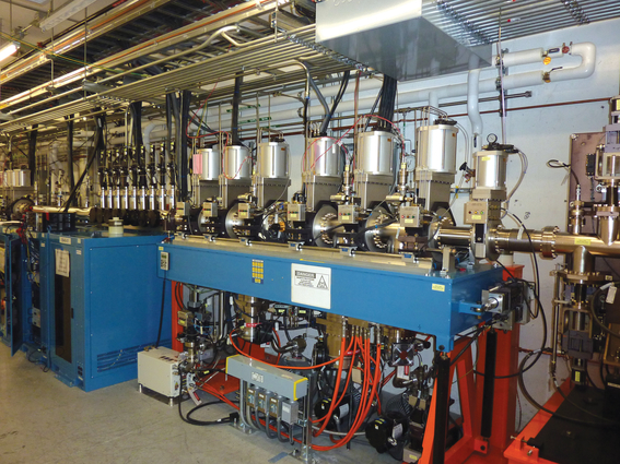
Livermore scientists designed and built the LCLS x-ray transport optics and diagnostics suite. One of the suite¿s instruments is the x-ray free-electron laser energy monitor, which won a 2010 R&D 100 Award for being one of the year's most important industrial innovations.
(Download Image)
Livermore scientists designed and built the LCLS x-ray transport optics and diagnostics suite. One of the suite¿s instruments is the x-ray free-electron laser energy monitor, which won a 2010 R&D 100 Award for being one of the year's most important industrial innovations.
Lab team helps measure femtosecond pulses of X-ray free electron
(Download Image)
Livermore scientists designed and built the LCLS x-ray transport optics and diagnostics suite. One of the suite¿s instruments is the x-ray free-electron laser energy monitor, which won a 2010 R&D 100 Award for being one of the year's most important industrial innovations.
An international team including three LLNL researchers have measured for the first time the spatial and temporal coherence of a single femtosecond X-ray pulse generated by the first hard X-ray free-electron laser (XFEL), the Linac Coherent Light Source (LCLS), at the SLAC National Accelerator Laboratory.
The LLNL team, which includes Regina Soufli, Stefan Hau-Riege and Monica Fernandez-Perea, developed and calibrated the optics in the Soft X-Ray Materials Science (SXR) beamline that were used to perform the experiment. The LLNL team also participated in the commissioning process of the SXR beamline, which included this experiment.
Soufli and Hau-Riege are members of a larger LLNL team which earlier, during LCLS construction, developed the LCLS optics and diagnostics that are used to filter and condition the XFEL beam before it enters the various beamline hutches (see the Science and Technology Review article ) .
The SXR beamline optics consist of three mirrors and two diffraction gratings and were made of super-polished silicon substrates coated with boron carbide. The optics had to be specially prepared to provide high reflectivity, maintain low stress and to preserve the coherence of the LCLS beam, which is crucial for the experiment, Soufli said. (Jeff Robinson and Sherry Baker from LLNL also participated in the SXR optics development).
Coherence is the fundamental property of light waves produced by laser sources. Combined with ultra short pulses, they yield insight into basic questions in physics through real-time observation and control of atomic scale structure and dynamics.
The intense, coherent and ultra short X-ray pulses from XFELS point to new insights into biology, condensed matter physics and atomic physics. The technique already has been used to image single viruses and pointed to new approaches to protein crystallography using nanocrystals.
Other research institutions include: Deutsches Elektronen-Synchroton DESY in Germany; National Research Nuclear University in Russia; University of California, Berkeley; SLAC National Accelerator Laboratory; the University of Melbourne; European XFEL GmbH of Germany; Universite Pierre et Marie Curie of France; ETH Zurich of Switzerland; Advanced Light Source, Lawrence Berkeley National Laboratory; Argonne National Laboratory; La Trobe University of Australia; and the University of Hamburg.
The research appears in the Sept. 30 issue of the journal, Physical Review Letters.
Oct. 20, 2011
[email protected]
925-422-9799
Science


Contact
Anne M Stark[email protected]
925-422-9799
Tags
Physical and Life SciencesScience
Featured Articles